Publications
Application of the Neuroplasticity Theory through the use of the Feldenkrais Method with a Canine with Traumatic Spinal Cord Injury: A Case Study
Oct. 2, 2012
Tammy Culpepper Wolfe, DPT, PT, CCRP, GCFP
The K9 Body Shop, PC, Arvada, CO
BACKGROUND AND PURPOSE
The Neuroplasticity Theory has gained widespread attention in the past several years in the medical and physical therapy professions. It states that, instead of being static, the central nervous system (CNS) can adapt its organization, structure, and function in response to changing internal and external stimulus.(1) There is current research and study focused on understanding neuroplasticity and how to take advantage of that plasticity. Although the theory is well documented, there is little documentation in the literature applying the theory to current treatment practices.(2) One of the least documented treatment methods is the Feldenkrais Method. Although the Feldenkrais Method has become a familiar term with clinicians in the physical therapy profession, there has been little clinical research to explain how it is performed and applied in patient care. While physical therapy on canines is based upon clinical evidence, only recently has there been research to test and describe the methodology of canine treatment regimen, practice, or efficacy.
The CNS has the capacity to adapt and alter its structure and function in response to a variety of internal and external pressures. This neural plasticity is the mechanism by which the CNS encodes experiences and learns new behaviors. It is also the mechanism by which the damaged CNS relearns lost behavior in response to rehabilitation.(3) Neuroplasticity includes the capacity of neurons throughout the CNS to change their structure and function in support of normal development and learning, as well as in response to injury or disease. Cortical maps can show reorganization in expanded synaptic connections among neurons and corresponding changes in function among those neurons. These maps were conventionally thought of as constant once an individual matured, but it is now understood that parts of them can change, expand, or shrink considerably.(4) Extended, skilled use of a body part causes its representation in the motor and somatosensory cortex to expand into surrounding areas. These changes occur relatively quickly (within hours), so it is thought that the changes probably depend on previously inactive, preexisting connections.4 In cases of injury, immobilization, or amputation, surrounding areas of the cerebral cortex take over the region of the affected part.4 After spinal cord injury, plastic changes occur at all levels of the CNS, including the cortex, other areas of the brain, and the spinal cord. These changes occur both rostral and caudal to the lesion.
It is thought that both spontaneous plasticity and activity-dependent plasticity can occur.(5) Spontaneous plasticity is thought to contribute to neurological return, but it may also have maladaptive effects, such as elevated muscle tone and pain. Activity-dependent plasticity occurs in response to afferent (sensory) input, causing adaptive neuronal changes. The mechanisms of activity-dependent plasticity appear to involve functional and structural changes at all levels of the CNS.8 On the behavioral level, there is recovery of sensory, motor, and autonomic function. At the spinal cord level, there may be normalization of reflexes and strengthening of motor-evoked potentials. Neuroanatomically, axonal and dendritic sprouting and even neurogenesis have been observed. In addition, on the cellular level, synaptic strengthening and up-regulation of neurotransmitters takes place.(6) It is activity-dependent plasticity that physical therapy intervention focuses on, and interventions are chosen that will develop the CNS in ways that will normalize function.
Although the Feldenkrais Method was being practiced before the Neuroplasticity Theory was developed, there has been little scientific explanation for how use of the method might achieve the excellent results that were being reported. The Feldenkrais Method was developed by Moshe Feldenkrais during the 1940s through the early 1980s. He was accomplished in Jujitsu and Judo and earned his degree in mechanical and electrical engineering and a Doctorate of Science in Engineering. After reinjuring an old soccer injury and deciding against knee surgery, he began developing what later became the Feldenkrais Method.(7) Since he chose to teach his method to the general public, the method developed in holistic, alternative medicine circles and has only recently been acknowledged as a legitimate form of treatment in main stream physical therapy. Due to the paths of the method development, there has been very little research published on the topic. What has been completed has primarily been focused on the Awareness Through Movement (ATM) aspect of the method. Intervention in this case study was based on the concepts of the other aspect of the method, functional integration (FI).
In ATM, the students (the term used by Moshe Feldenkrais, instead of “patients”) are verbally instructed to move in a series of very specific ways, one movement building upon the previous sequence of movements; FI is an intensive, individual-specific manual technique. In FI, the teacher (practitioner) uses various manual techniques to promote changes in the CNS by communicating to the student how they habitually move, and then offers different movement options for better efficiency, coordination, and fluidity. In the process, the CNS develops new functional motor patterns and new patterns of movement emerge. At times, those new patterns emerge immediately and permanently, and at other times, the patterns and changes may occur over several days after a lesson. The slower changes is seen several times over weeks or months, new movement patterns may emerge at any time during that period of lessons as one lesson builds upon another.
The changes that take place can be explained by the Neuroplasticity Theory, given the assumption that the activity-dependent plasticity occurs during the FI lesson. Activity-dependent plasticity depends upon sensory input from an external stimulus. During an FI lesson, the practitioner gives various types of manual sensory input to the student to allow them to explore and learn new possibilities of functional movement, using their bodies in ways that are unfamiliar or have been forgotten because of injury or illness. The manual techniques facilitate learning of new connections throughout the body and result in movement patterns that are more efficient, comfortable, and functional.
Although an FI lesson uses some manual skills already mastered by a physical therapist, the intention of the practitioner is more instructive than corrective in nature. Through kinetic rapport, the student learns how to reorganize his body posturing and movements, including his limitations, in new and more effective ways. The areas in which he operates effectively and comfortably then begin to expand into other functions not previously achievable.(8) According to the Neuroplasticity Theory, this unusual, purposeful and functional sensory input causes the CNS to form new connections at every level.
As an example of how standard PT techniques can be used in a Feldenkrais Method is described here. A joint may be mobilized during an FI lesson, but it will not be mobilized as an isolated action. It will always be mobilized as part of a functional movement, in relationship to the rest of the body’s whole movement pattern, and in relationship to the changes taking place in the whole body at the time. For example, thoracic vertebra T8 may be mobilized as part of a dynamic “reaching across midline” movement, instead of statically without the student’s input. Information is given in various situations and settings for the purpose of allowing neuroplasticity to take place in a broader spectrum instead of only in specific circumstances.1
This case study was chosen to demonstrate the Neuroplasticity Theory because FI was done in various positions and ways to assist the patient in re-learning a variety of functional patterns in several body positions.
CASE DESCRIPTION
The patient was a two-year-old, active, female Shiba Inu, without prior health conditions. The 26-pound dog was in a car during an automobile accident and was thrown head-first into the windshield upon impact. After receiving medical care in an emergency veterinary hospital, she arrived for physical therapy 10 days following the accident.
The patient was being carried by her owner upon arrival. When she was placed on the rug, she had difficulty maintaining a lying position while watching the others in the room without losing her balance.
Examination
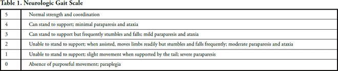
The patient was bright, alert, and responsive (BAR), and her pulse and heart rate were normal and regular. She was non-ambulatory or grade 1 on the neurologic gait scale (Table 1 – above).(9) She required two people to transfer from sternal to standing and to maintain a standing position. She was able to transfer and maintain sitting with assistance of one person using both hands to assist and stabilize her in that position. She was able to independently transfer from right lateral recumbency to sternal, but she required a one-person assist to transfer from left lateral recumbency to sternal. Her posture in standing was Schiff-Sherrington positioning bilaterally with upper motor neuron signs and extensor tone in forelimbs (FLs) and lower motor neuron signs and flexor tone in the hind limbs (HLs).(10) Cervical right rotation was 50% of normal. All other passive range of motion was normal, as defined by Jaegger et al.(11) Muscle spasms were palpable in the epaxial muscles bilaterally from the cervical through the lumbar spine and in the bilateral external obliques. Proprioception reflexes were assessed in standing position and were delayed in the left FL and left HL and absent in the right FL and right HL. Patellar reflexes were 3+ bilaterally. Flexor withdrawal reflexes were intact bilaterally and extensor patterning was present only on the right HL. Both tests were done in lateral recumbency. There was no pain response on examination. Deep pain sensation when tested with hemostats was intact in all 4 extremities. She was able to initiate voluntary movement in all 4 extremities.
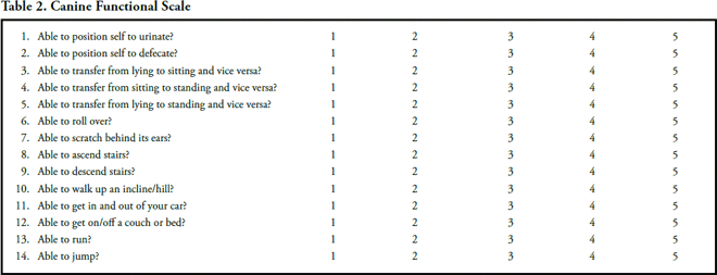
Her owner’s goals were to maximize his dog’s functional independence and to reach the highest quality of life possible. He stressed his desire for her to walk independently and hoped for normal bowel and bladder control, which she did not have at the time. She was scored as a 14/56 on the Canine Functional Scale (Table 2 – above).(12) Her prognosis for reaching the goals was good, based on the fact that her deep pain sensation was intact and that she had voluntary muscle contraction in all extremities.(13)
A diagnosis of tetraparesis with possible cervical disc herniation was made, based upon examination findings. The dog clearly needed to form new neurologic connections and re-learn basic functional movements, coordination, and balance to be able to function normally again. Therefore, she was an excellent candidate.
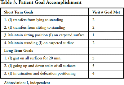
The plan of care was to see the patient on a weekly basis for FI and implementation of a progressive home care program. Treatments would be spread further apart, based upon the patient’s progress and the client’s economic constraints. Underwater treadmill exercise for strengthening and balance training(14) was to be added when the patient was able to ambulate in water with assistance of one person. Short-term goals were: (1) independent transfers from lying and sitting to standing; (2) maintain sitting position without assistance on carpeted surface; and (3) maintain standing without assistance on carpeted surface. Long-term goals were: (1) independent gait on all surfaces for 20 minutes; (2) independence going up and down stairs of all surfaces; and (3) independence in urination and defecation positioning (Table 3 – above).
INTERVENTION
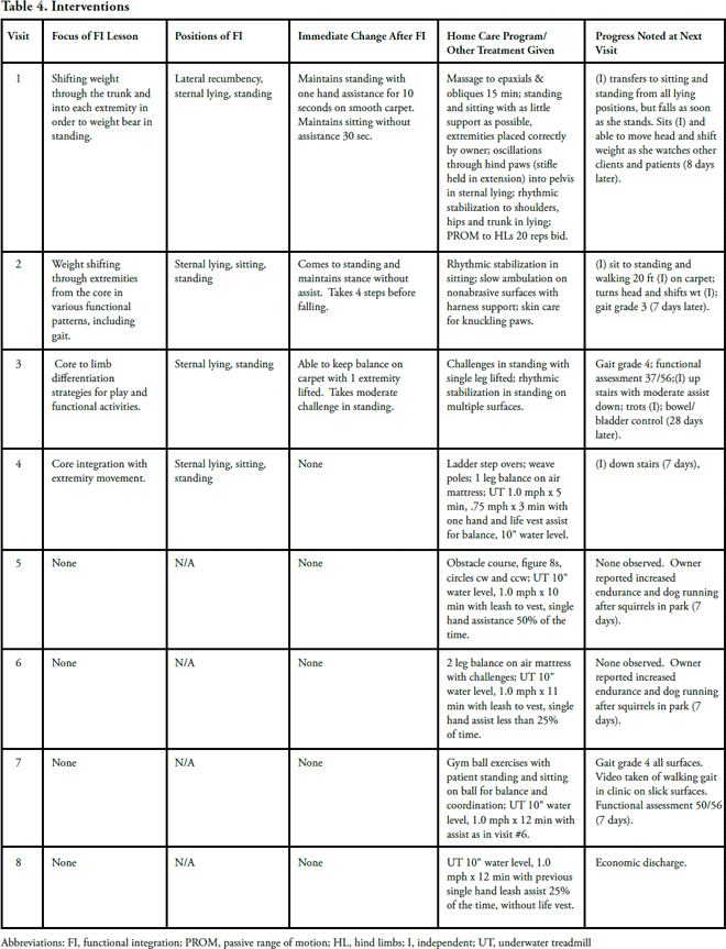
Because of the nature of FI, it would be impractical and nearly impossible to document exactly what was done in a 20 to 30 minute FI lesson. Although each Feldenkrais Practitioner is trained in a 4-year program and spends 800 to 1000 hours in class, each practitioner has an individualized approach to his or her performance of a lesson. However, any practitioner who was provided with specific functional movements to facilitate, would structure the lesson in a similar manner. Table 4 (below) illustrates the intervention in its logical order.
The patient was seen for a total of 8 visits over a period of 11 weeks. Because of the client’s economic situation, the patient received FI as part of the treatment for only the first 4 visits (Table 4). All home care exercises were to be done twice daily. Repetitions were determined by the dog’s continued willingness to perform the exercise. The owner was given written instructions and pictures to assist him in performing the home care program. The exercises given are well known in the canine physical therapy profession. Rationale for home exercise progression was based on the dog’s ability to master the previous exercises. The rationale for FI focus was based upon the functional activity and movement patterns that the dog needed to re-learn in order to return to normal activity. The sequence was based upon simple to more complex activities and movements and also based upon the movements that the dog was able and willing to learn in any given lesson.
OUTCOMES
Animal physical therapy is a relatively new specialty field with little standardization of any outcome forms, measurements, or tests. There are 4 gait or lameness scales the author is familiar with, two pain scales, several functional scales, and two body composition scales–none of which have any studies of reliability or validity. However, with the given lameness grading and functional assessment scales used in the case study, progress toward the client’s goals can be identified. Both scales are easy to use and have objective guidelines in which to assess the patient’s status. Improved functional activities were documented, both immediately after FI and between visits to physical therapy. The lameness grade improved from zero to 4 out of 5 possible stages. The assessment score improved from 14 to 50 out of 56 possible points. Despite the lack of evidence of reliability or validity in these measures, there were observable improvements in the functional status of the patient as a result of physical therapy intervention.
DISCUSSION
The purpose of this case was to apply the Neuroplasticity Theory by use of FI as a mode of rehabilitation of a canine with a traumatic spinal cord injury. While it is impossible to know what changes took place structurally and chemically in the CNS of this dog during the 11 weeks of physical therapy intervention, it is possible to observe the outcomes of this intervention. At the time of evaluation, the patient exhibited signs of significant CNS disruption and injury, resulting in Schiff-Sherrington posturing and severely limited functional mobility. Following the FI sessions, there were immediate changes in functional patterns of movement, such as the ability to maintain sitting and standing balance and the ability to take independent steps. Drawing on the assumptions of the Neuroplasticity Theory, one conclusion may be that activity-dependent neuroplasticity took place in the CNS during the FI lessons. Research has shown a high correlation between early functional training with appropriate sensory input and improved walking function;(15) however, it is impossible to isolate the contribution of the FI versus the balance and strengthening exercises.
This study is one of many studies needed in the areas of neuroplasticity and rehabilitation. One of the questions that the study leaves unanswered is if FI is as effective in changing the CNS and improving functional movement patterns as other currently used physical therapy techniques and modalities. As in many canine neurological injuries, it remains unknown as to how much of the dog’s recovery might have been due to spontaneous plasticity. Since the original spinal cord injury was not quantified by advanced diagnostics, the structural recovery taking place in the CNS was impossible to assess.
To assist in assessing the actual change taking place during the treatment process, standardization of reliable and valid measurement tools represents another aspect of this case study that requires further research. This case study demonstrates the need for more research and creative thinking concerning how physical therapists can develop more effective treatment techniques while using our ever-growing understanding of neuroplasticity of the CNS after injury and throughout the rehabilitation process.
REFERENCES
- Young JA, Tolentino M. Neuroplasticity and its applications for rehabilitation. Am J Ther. 2010;18:70-80.
- Tansey KE. Neural plasticity and locomotor recovery after spinal cord injury. Phys Med Rehabil. 2010;12:S220-S226.
- Kleim J, Jones TA. Principles of experience-dependant neural plasticity: Implications for rehabilitation after brain damage. J Speech Lang Hear Res. 2008;S1:S225-S239.
- Pascual-Leone A, Amedi A, Fregni F, Merabet LB. The plastic human brain cortex. Ann Rev Neurosci. 2005;28:377-401.
- Dunlop AS. Activity-dependent plasticity: Implications for recovery after spinal cord injury. Trends Neurosci. 2008;33:410-418.
- Lynskey JV, Belander A, Jung R. Activity-dependent plasticity in spinal cord injury. J Rehabil Res Dev. 2008;45:229-240.
- The Feldenkrais Method® of Somatic Education. The Biography of Moshe Feldenkrais. Accessed on September 12, 2011.
- Feldenkrais Movement Institute. The Feldenkrais Method® in Clinical Applications. http://www.feldenkraisinstitute.org
Accessed on September 12, 2011. - Millis D, Taylor R, Levine D, et al. Introduction to Canine Rehabilitation. Knoxville, TN: University of Tennessee; 2002.
- Schadt JC, Barnes CD. Motoneuron membrane changes associated with spinal shock and the Schiff-Sherrington phenomenon. Brain Res. 1980;201(2):373-383.
- Jaegger G, Marcellin-Little DJ, Levine D. Reliability of goniometry in Labrador Retrievers. Am J Vet Res. 2002;63:979-986.
- Adrian C. Alameda East Veterinary Hospital Sports Medicine Rehabilitation Dept. Functional Mobility Scale; 2001.
- Dhupa S, Glickman NW, Waters DJ. Functional outcome in dogs after surgical treatment of caudal lumbar intervertebral disc herniation. J Am Amin Hosp Assoc. 1999;35:323-331.
- Stevens S, Morgan DW. Underwater treadmill training in adults with incomplete spinal cord injuries. J Rehabil Res Dev. 2010;47(7):vii-x.
- Deitz V. Neuronal plasticity after a human spinal cord injury: positive and negative effects. Exp Neurol. 2012;235(1):110-115.
Taken from the Orthopaedic Practice Vol. 24;4:12.